


Dangerous fascination
Комплексний, індивідуальний, анонімний підхід до лікування.
There are attractive marks on the body, which are all called moles, have long been considered the secret of ladies’ charm. However this component of the unique female charm can turn into serious health problems for its owner.
Moles are an everyday name for congenital and acquired pigmented nevi on the skin, often in the form of a dark brown spot or pea. In medicine, there is a term «nevus», that in general means the pigment nevi of the skin.
Why do moles appear?
Nevus (congenital or acquired) is formed on the skin by pigment cells that produce melanin-melanocytes, which are normally located between the two layers of the skin: the epidermis (the top layer) and the dermis (the inner layer), and has already existed there at birth.
Methods of removing moles
There are two categories of indications for removing moles — cosmetic and oncological. Depending on this, the method of their removal is determined. But only specialist must make a choice.
Methods of removing moles in the medical center «Dermatology Plus»
- Laser – a laser beam is used, penetrating into the skin strictly to a predetermined depth.
- cryodestruction (liquid nitrogen) – removal by low-temperature controlled exposure of liquid nitrogen, whereby there is a mortification (frostbite) of mole;
- radiowave apparatus Surgetron: relative painlessness of the procedure;
- rapid healing;
- no burns after removal;
- impact directly on the neoplasm without damaging the adjacent skin areas;
- it is possible to carry out of histological analysis of biological material;
- bloodless procedure;
- carrying out theprocedure on tanned or swarthy skin;
- minimal risk of inflammation, wound abscess, appearance of pigmental spots
- and other consequences;
- the procedure does not require rehabilitation and hospitalization.
- electrocoagulation – is using a high-frequency discharge of an electric current, which plays the role of a scalpel, with the help of it micro-cuts are carried out and the nevus is cut off in small layers;
- Jett Plazma Medical apparatus
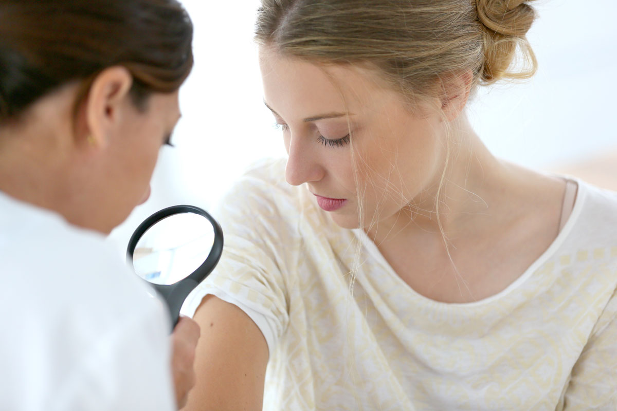
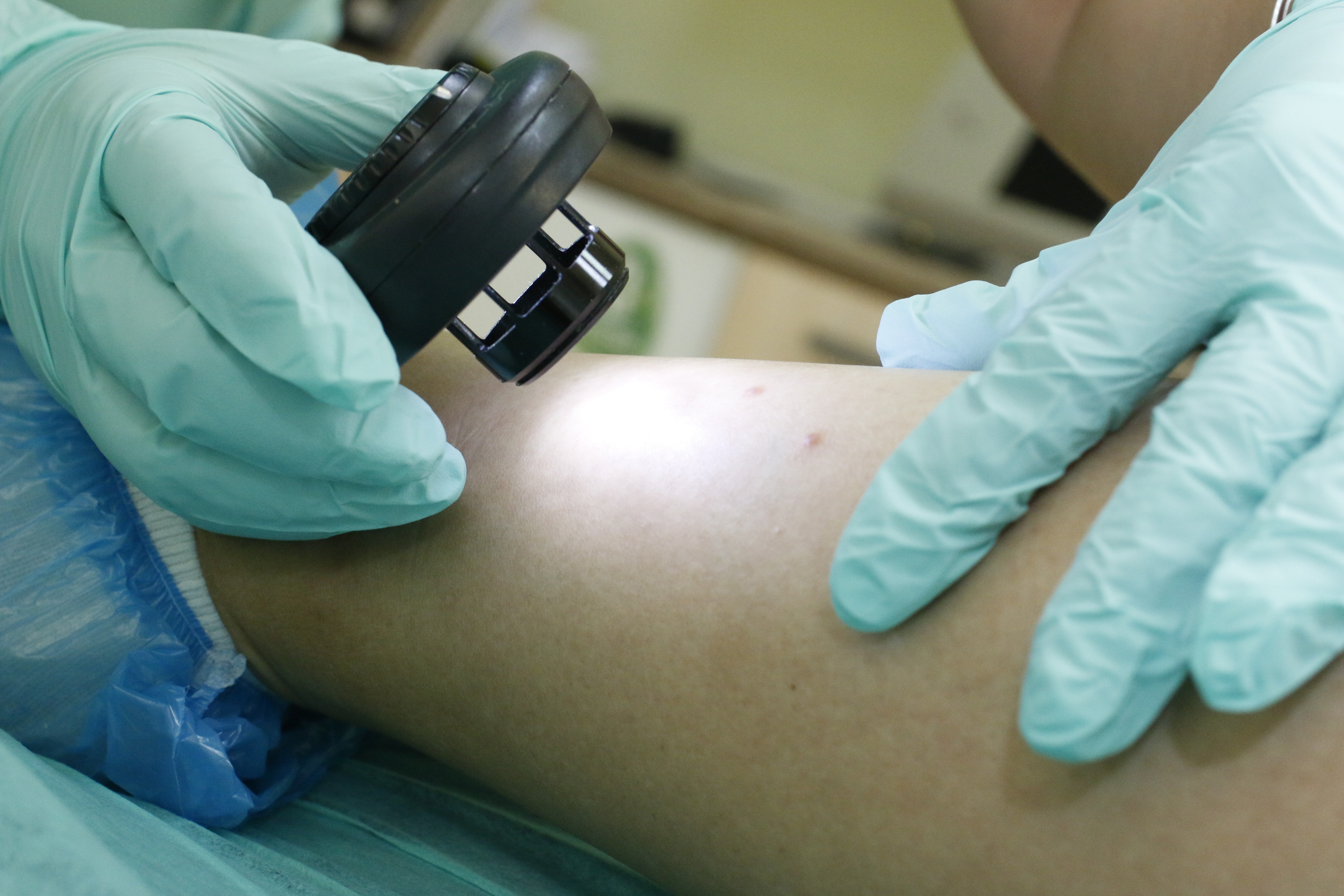
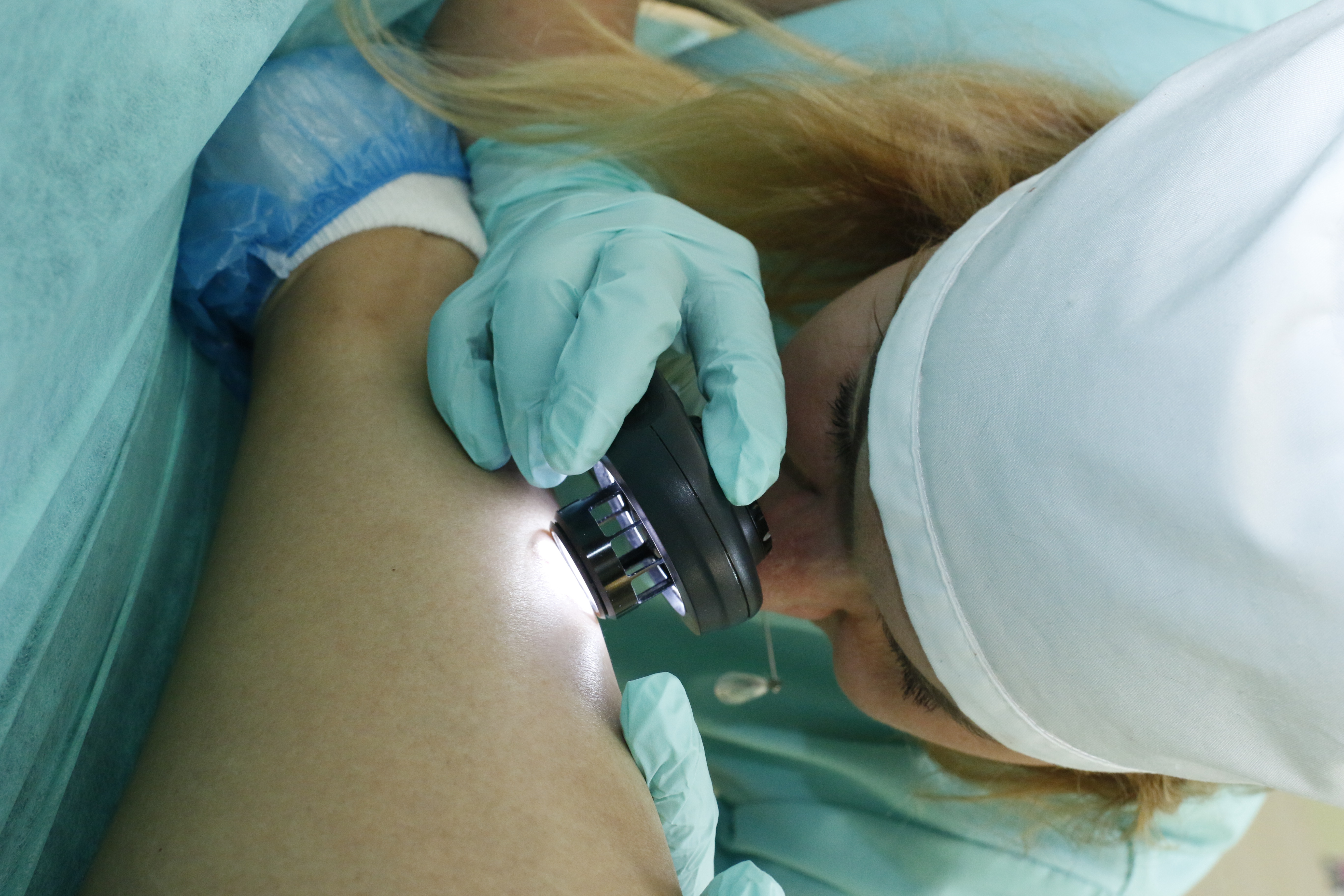
Specialist has conversation with the patient in order to correctly give the diagnosis finding out congenital or acquired pigmented is this neoplasm, what form had it at first, were some changes and for what period of time happened and what is the cause and also whether already treatment has carried out of nevus and how. In addition, the doctor is sure to do examination of the neoplasm, specifying its color, shape, size, etc.
The standard methods of diagnosis of any pigment neoplasms of the skin including melanoma comprehend histological examination (the study of tissue samples taken from the patient, under a microscope to study the structure. The material for the study is obtained as follows: do a smear-imprint from the surface of the pigment formation (in the presence of cracks and bleeding on it) and examined it. The results in this case
are usually ready the next day.
Sometimes, immediately after the examination, the suspicious neoplasm is completely removed with minimal indentation (3-5 mm) from its edges for further histological examination. The procedure is сarried out under local anesthesia. Results can be obtained in a few days. It is important to note that all of these actions should be carried out only in specialized oncological institutions.

Snezhko Svetlana
Dermato-oncologist. Cosmetologist. Dermatovenerologist.
Association of Dermatovenereologists and Cosmetologists of Ukraine
Working hours
Monday- Friday: 9.00-19.00
Saturday / Sunday: day off