


The soft fibroma (papillomas, acrochordon). Seborrheic keratomas. Keratoacanthoma.

Сomplex, Individual, Anonymous Approach To Treatment.
Soft fibroids (papillomas, acrochords).
Mostly papillomas and soft fibromas occur in middle-aged people, often the background for their appearance are disorders of the endocrine system.
Clinical implications.
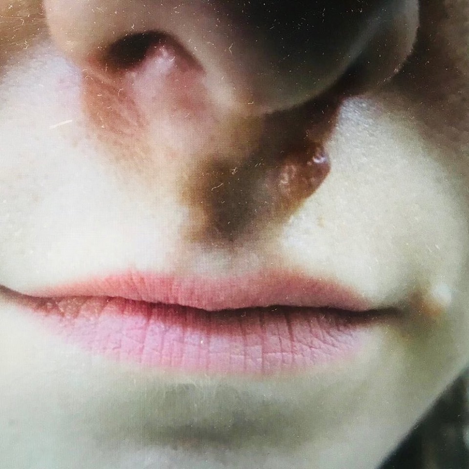
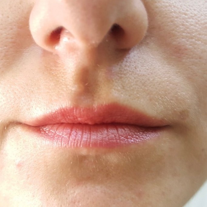
Papillomas usually have the color of the surrounding skin is slightly darker than it, their size is often 0.5-5 mm.
Papillomas are characterized by slow growth, but sometimes, for example, during pregnancy, they progress quickly.
Differential diagnosis.
Threadlike warts are located on the fingers of the hands, reminding a cutaneous horn. Seborrheic keratomas usually have a larger size, dark color, warty surface. Neurofibromas are quite large, often localized on the skin of the back and are hereditary; single elements are not an indicator of systemic disease.
Seborrheic keratomas
Seborrheic keratomas are a common type of epithelial tumors. Mostly they occur in old age. The number of keratomas can be different from single elements to several hundred, especially often keratomas affect areas of oily skin. The abundance of seborrheic keratomas can sometimes be a manifestation of borderline cancer.
Clinical implications.
Seborrheic keratomas are most often localized on the face, neck, scalp, back and upper half of the chest, less often on the forearms, shins and lower half of the trunk. Usually their diameter does not exceed 1 cm, rarely reaches 3 cm or more. Is dominated by eruptions of yellow, brown, sometimes black. Keratomas have an oval shape with a warty surface, slightly rise above the level of the skin, covered with a thin fatty film, for which they received their name. The characteristic symptoms are a white, brown, or black tube (pseudologue cysts).
Stage of development.
At an early stage, small papules practically do not rise above the skin surface and are often pigmented. Their surface is scrobiculated. In the later stages of development, keratomas are transformed into warty plaques that are above the surrounding skin in the form of a nail head.
Differential diagnosis.
Pigment nevi exist for a long time, have a smooth surface and elastic consistency. Flat warts are observed more often in children and young people. They appear suddenly and often in large clusters. Melanoma is rare and usually characterized by rapid growth with induration at the base.
Keratoacanthoma
Keratoacanthoma is a fast-growing epithelial tumor with a massive cluster of horn masses, which are located in the center. Most freaquently keratoacanthomas affect the face and upper limbs.
Clinical implications.
The tumor resembles squamous cell carcinoma, but as opposed to it often regresses spontaneously in 6-9 months after onset. Although this tumor is not usually regarded as a primary malignant neoplasm, the incidence of malignancy of keratoacanthoma, according to some authors, reaches 60%. In this case, the main factor of malignancy is ligering bleeding of the ulcer base after rejection of the horn masses and the appearance of an induration at the base of the element.
The most common type of elements is solitary. Less common are atypical keratoacanthomas (giant, subungual, centrifugum marginatum, multinodular, etc.), appearing constantly during the patient’s life. They do not have an appointed localization.
There are 3 stages of keratoacanthoma development: growth stage, stabilization stage and regression stage. During the growth stage small papule ,which suddenly appeared, acquires a rich red color, its diameter within a few weeks reaches 1-2 cm.
At the same time, there are no subjective sensations. During the stabilization of the keratoacanthoma, the growth of the element stops and a cup is formed, filled with gray horn masses. At the stage of regression, the horn masses are separated and the tumor completely regresses, leaving a subtle atrophic cicatrix.
BENIGN TUMORS
Treatment of widespread benign tumors is often surgical. Radiation therapy with soft x-rays is also used. Physical therapy is aimed at destruction of the tumor cells (dermodestruction methods) and their removal (coagulation techniques). Some seborrheic warts for cosmetic reasons or with the threat of malignancy are subjected to cryodestruction with liquid nitrogen or destroyed by other methods. Treatment of keratoacanthoma represents the excision of mass within the unaffected tissue. According to some data, satisfactory results are given by local application of cytotoxic agents (Methotrexate, etc.) and x-ray therapy (15-30 Gy) of the tumors treatment.
Treatment of skin nevus is also surgical, depending on its size, site and clinical implications. A large face nevus, lading to aesthetic breach, is excised with a single stage local tissue rearrangement or a free autodermic grafting or a stepped excision is used. Nevus (even small) is subjected to permanent injury (by collar, glasses, comb, etc.) is removed. When tumor symptoms appear, additional radioisotope diagnostics is required, which allows to determine the benign or malignant nature of the process.
By maintaining the benign nature of the nevus, it is necessary to excise it, while the boundaries of the operation should be expanded. In recent years, cryodestruction has been widely used for the treatment of nevi.


Snezhko Svetlana
Dermato-oncologist. Cosmetologist. Dermatovenerologist.
Association of Dermatovenereologists and Cosmetologists of Ukraine
Working hours
Monday- Friday: 9.00-19.00
Saturday / Sunday: day off